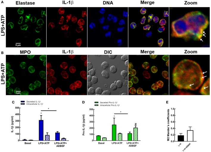
Figure 6.
Role of serine proteases in interleukin-1β (IL-1β) secretion. Colocalization of IL-1β with elastase (A) or myeloperoxidase (MPO) (B). Neutrophils were stimulated with LPS and 2 h later were treated with ATP. At 3.5 h post-LPS stimulation, cells were fixed, permeabilized, and stained with specific antibodies anti-IL-1β (red) and elastase [(A); green] or MPO [(B); green]. Images were acquired with a confocal microscope. Arrows indicate examples of compartments with IL-1β and elastase or MPO overlapping signals. Concentrations of IL-1β (C) and pro-IL-1β (D) in culture supernatants and cell pellets of neutrophils stimulated or not for 5 h with LPS + ATP in the absence or presence of AEBSF (1 mM) added 1 h post-LPS stimulation. Data represent the mean ± SEM of four experiments performed in duplicate. Two-way ANOVA followed by Bonferroni’s multiple comparisons test. * and # p < 0.05 vs. their respective basals or between LPS + ATP-treated or not with AEBSF. (E) Manders’ colocalization coefficients of IL-1β/LC3B for IL-1β (M2) from images of neutrophils stimulated 4 h with LPS + ATP and treated or not with AEBSF (1 mM) 1 h after LPS stimulation. Values were derived from five independent experiments with at least 60 cells analyzed per experiment. Results were evaluated by Mann–Whitney test analysis and differences were not statistically significant.

