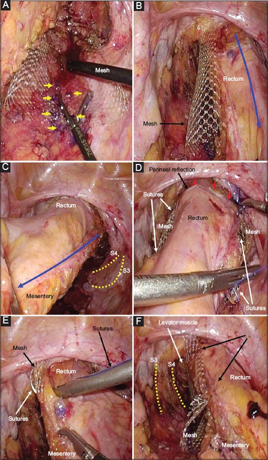
Figure 6.

(A) Mesh is fixed to the pre-sacrococcygeal fascia (arrows) using absorbable tacks. (B, C) The mobilized rectum is lifted cranially to the promontorium (blue arrow). Pelvic nerves (dotted lines) are preserved. (D-F) The seromuscular layer of the rectal wall is sutured to the mesh using a small number of interrupted non-absorbable polypropylene sutures. Pelvic nerves (dotted lines) are preserved. The anterior wall is elevated (red arrows)
