Figure 1.

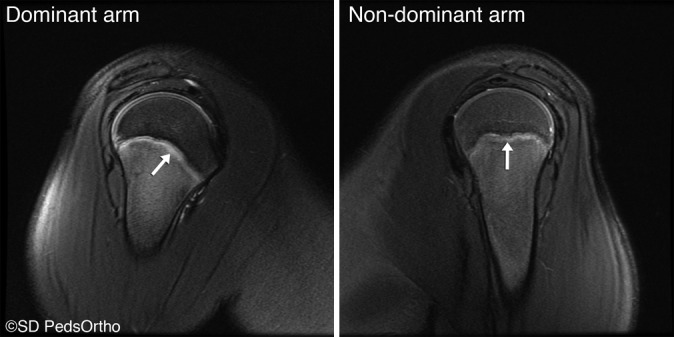
Sagittal T2-weighted fat-suppressed image demonstrating periphyseal edema. (Reprinted with permission from San Diego Pediatric Orthopedics.)

Sagittal T2-weighted fat-suppressed image demonstrating periphyseal edema. (Reprinted with permission from San Diego Pediatric Orthopedics.)