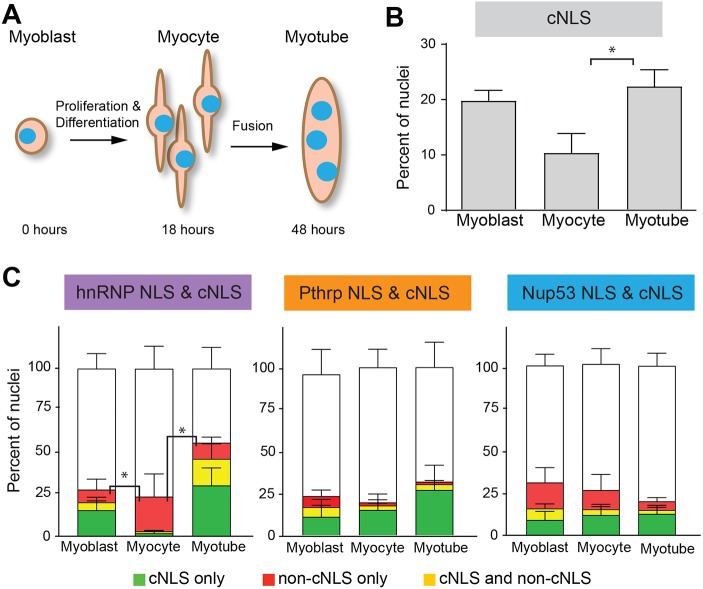
Fig. 6.
Nuclear import varies during myogenesis in vitro. (A) Pure cultures of primary myoblasts were differentiated to form myocytes or myotubes and the in vitro nuclear import assay was performed. (B) The percentage of cNLS-positive nuclei was high in myoblasts (20%), dropped dramatically in myocytes (9%) and then rose again to pre-differentiation levels in myotubes (23%). (C) The in vitro import assay was performed using cNLS and non-cNLS import reporters to compare the relative import of the cNLS and non-cNLSs at different stages of myogenesis. For each pair of NLS reporters examined, the proportions of nuclei importing the non-cNLS only or both the cNLS and non-cNLS were compared, as in Fig. 3C. The percentage of hnRNP NLS import differed significantly across differentiation. The hnRNP reporter displayed relatively low import in the myoblast and myotube stages but relatively high import in the myocyte stage. All data are mean±s.e.m.; *P<0.05, comparisons by ANOVA with Tukey correction for multiple comparisons; n=3 independent experiments, each with ∼200 nuclei per differentiation stage.

