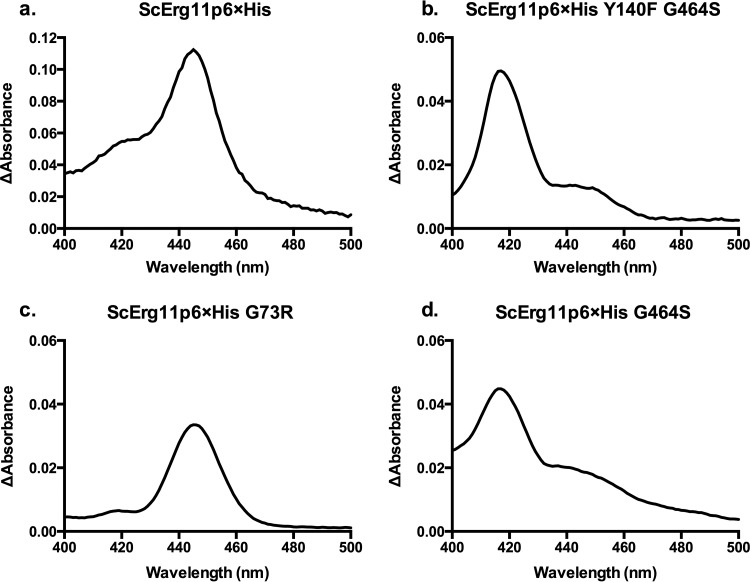
FIG 2.
Wild-type and mutant ScErg11p6×His carbon monoxide difference spectra. Difference spectra are shown for the wild-type ScErg11p6×His protein (a) and the Y140F G464S (b), G73R (as a representative of the G73W/R/E mutants, as all 3 profiles were similar) (c), and G464S (d) mutants. The difference spectra were obtained by using equal concentrations of the ScErg11p6×His protein in a control sample, which was reduced by sodium dithionite, and a test sample, which was reduced by sodium dithionite after the solution was saturated with carbon monoxide. The peak at 445 nm represents functional CYP450, and the peak at 417 nm represents nonfunctional CYP450.

