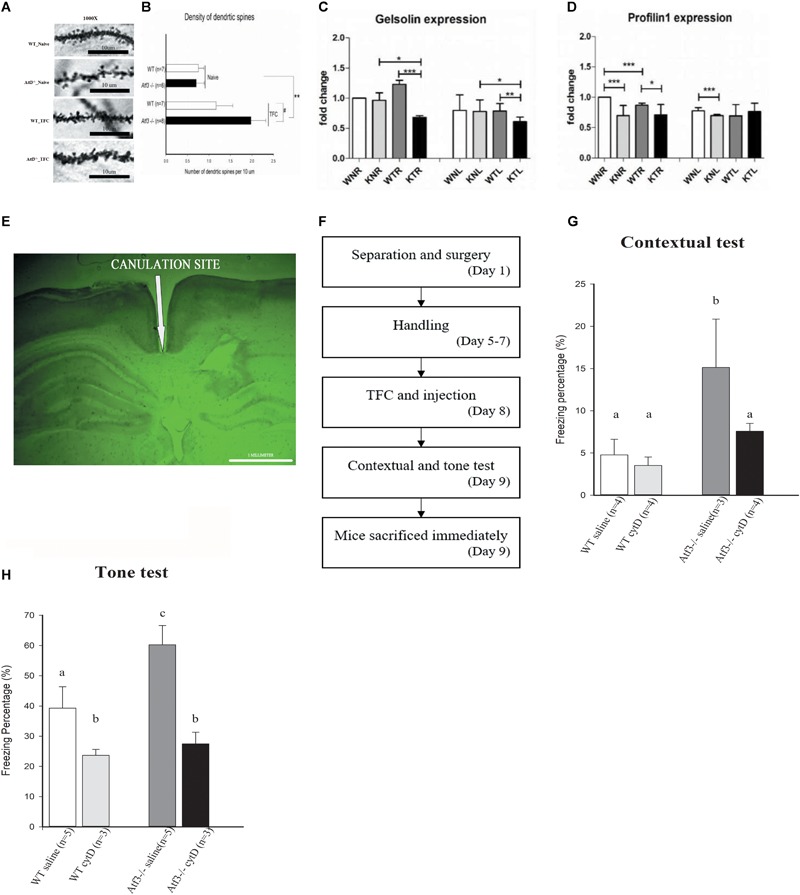
FIGURE 5.

Loss of ATF3 increases dendritic spine density in the hippocampal region after retrieval of contextual trace fear memory. Inhibition of actin polymerization through intracranial infusion of cytochalasin D reversed the phenotype of Atf3-/- mice. Microscopic graphs of dendritic spines in the hippocampal CA1 region (A). Quantification of spine density of basal dendrites measured after retrieval of contextual memory for the Atf3-/- mice and their wild-type littermates with or without trace fear conditioning (B). mRNA expression levels of Gelsolin (C) and Profilin 1 (D) in wild-type and Atf3-/- mice before and after training (n = 3 for each sample; W, wild type; K, knockout; N, naïve; T, trained groups; L, left hippocampus; R, right hippocampus). Position of cannula placement (AP: –2.18 mm from the bregma, DV: 1.8 mm. (E). Timeline for intracranial infusion and fear conditioning (F). Freezing percentage to context (n = 4: WT saline, 4: WT treated with drug, 3: KO saline and 4: KO treated with drug) (G) and to tone (n = 5: WT saline, 3: WT treated with drug, 5: KO saline and 3: KO treated with drug) (H) after injection of cytochalasin D (cytD). The results shown in the figure are plotted as mean ± SE and statistically tested with one-way ANOVA followed by Tukey test and set a confidence level of 99% (p < 0.001). a, b, and c indicate significant difference among groups with different letters (P < 0.05).
