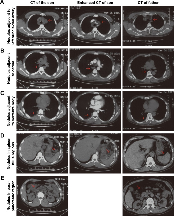
Figure 2.
The CT scan and enhanced CT scan of the son and CT scan of the father.
Notes: Both CT scan methods confirmed the plexiform lesions in the subclavian artery region (A), carina region (B), vertebra body region (C), spleen hilus regions (D), and para-pancreatic region (E), which could not be enhanced by tracer and were unique in the son while absent in the father.
Abbreviation: CT, computed tomography.

