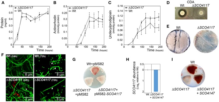
Figure 2.
Phenotypical analyses of the ΔSCO4117 mutant. (A) Growth curve. (B) Actinorhodin production. (C) Undecylprodigiosin production. (D) CDA production. (E) Macroscopic view of sporulation (gray color) in the ΔSCO4117 mutant compared to the wild strain in GYM plates at 85 h. (F) Confocal laser fluorescence microscopy pictures (SYTO9-PI staining) of the ΔSCO4117 mutant illustrating delay in MII differentiation (24 h) and sporulation (72 h) compared to the S. coelicolor M145 wild-type strain (GYM plates). Arrows indicate spore chains; the asterisk indicates the discontinuities characterizing the MI compartmentalized hyphae. (G) Macroscopic view of antibiotic production (red color) in the wild-type strain, the ΔSCO4117 mutant harboring plasmid pMS82 and the ΔSCO4117 mutant harboring plasmid pMS82-SCO4117, all of them grown in MM plates at 5 days. (H) SCO4117 transcript abundance in the ΔSCO4117 mutant harboring pMS82-SCO4117 (complemented mutant) compared to the wild-type strain in GYM plates at 17 h. (I) Macroscopic view of antibiotic production (red color) in the ΔSCO4117 restored mutant in MM plates at 5 days.

