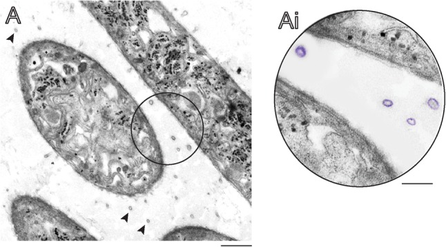
FIGURE 1.

Longitudinal and cross sections of Cylindrospermopsis raciborskii growing in control conditions seen by transmission electron microscopy (TEM). (A) Several extracellular membrane vesicles (indicated by arrowheads in A and highlighted in purple in Ai) are observed around cultured cyanobacterial cells.
