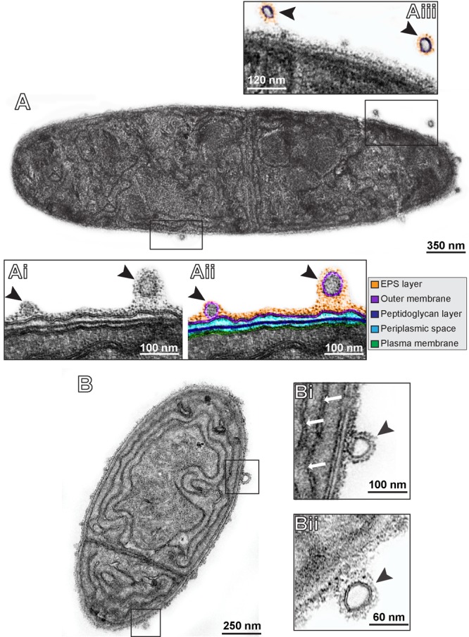
FIGURE 2.

Representative electron micrographs of C. raciborskii cells under control culture conditions (A,B). In (Ai,Aii), the cell envelope is seen in high magnification. This structure is composed of two bilayered membranes: the inner or plasma membrane (highlighted in green) and the outer membrane (highlighted in purple) that encloses the periplasmic space (light blue) with a thin peptidoglycan layer (dark blue). Note the presence of OMVs (arrowheads) with typical trilaminar structure cleary budding off from the outer membrane. Secreted vesicles frequently exhibited an external amorphous material (extracellular polymeric substances – EPS) as observed on the surface of the cell envelope (highlighted in orange in Aii,Aiii). Thylacoid membranes are indicated by white arrows in (Bi).
