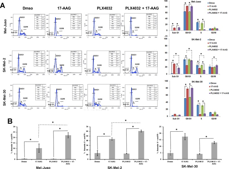
Fig 6. Inhibitory effects of 17-AAG on cell cycle arrest and apoptosis of PLX4032 pretreated human melanoma cells.
(A) (left panel) Cell cycle distribution was determined by flow cytometry, on Mel-Juso, SK-Mel-2 and SK-Mel-30 cells pre-incubated with PLX4032 (2μM) or vehicle (DMSO) for 24h with subsequent incubation with or without 17-AAG (1μM) for an additional 72h. Cell cycle profiles were obtained by cell population-based DNA content analysis by flow cytometry (propidium iodide staining) of treated and untreated cells. (Right panel) Graphical representation of the data. The experiment in (left panel) was performed in triplicate and the percent of cells in Sub G1, G0/G1, S, and G2/M was quantified. Data (mean ± SD, n = 3); *P<0.01. (B) Effect of apoptosis was determined by flow cytometry on human melanoma cells (Mel-Juso, SK-Mel-2, SK-Mel-30) pre-incubated with PLX4032 (2μM) or vehicle (DMSO) for 24h, with subsequent incubation with or without 17-AAG (1μM) for an additional 72h. The data is a mean of percentage of Annexin V-positive and PI-negative cell population from three independent experiments. *P<0.01.

