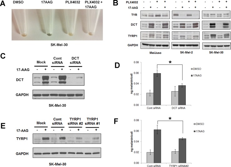
Fig 7. HSP90 inhibition by 17AAG induces pigmentation in human melanoma cells.
(A) SK-Mel-30 human melanoma cells pretreated with or without PLX4032 were further treated with 17-AAG or vehicle for 72h, and then harvested and spun down. Data shows the cell pellet color.17-AAG alone induced pigmentation in human melanoma cells. (B) Human melanoma cells (Mel-Juso, A375, SK-Mel-30 and SK-Mel-2) were pre-incubated with PLX4032 (2μM) or vehicle (DMSO) for 24h and subsequently incubated with or without 17-AAG (1μM) for 48h. Cell lysates were collected and studied for the expression of TYR, DCT and TYRP1 proteins by western blot. Data show 17AAG induced DCT and TYRP1 protein expression regardless of PLX4032 treatments whereas TYR was either unchanged (Mel-Juso, SK-Mel-30) or decreased (SK-Mel-2). (C) SK-mel-30 melanoma cells transfected with a control siRNA or DCT siRNA for 24h were incubated with or without 17-AAG for 48h. DCT knockdown was confirmed by western blot. (D) SK-mel-30 melanoma cells transfected with DCT siRNA show significant reduction in melanin content in 17AAG-treated cells in comparison to control siRNA treated cells. Data are represented as percentage of control and SD, measured from three independent experiments. *P < 0.05 vs. control siRNA + 17-AAG. (E) SK-Mel-30 melanoma cells transfected with a control siRNA or 2 different TYRP1 siRNAs (#1, #2) for 24h. They were further treated with or without 17-AAG for 48h. Knockdown of TYRP1 expression was confirmed by collecting cell lysates and running a western blot. (F) SK-Mel-30 melanoma cells transfected with TYRP1 siRNA #2 shows significant reduction in melanin content in 17AAG treated cells in comparison to control siRNA. Data are represented as percentage of control and SD, measured from three independent experiments. *P < 0.05 vs. control siRNA + 17-AAG.

