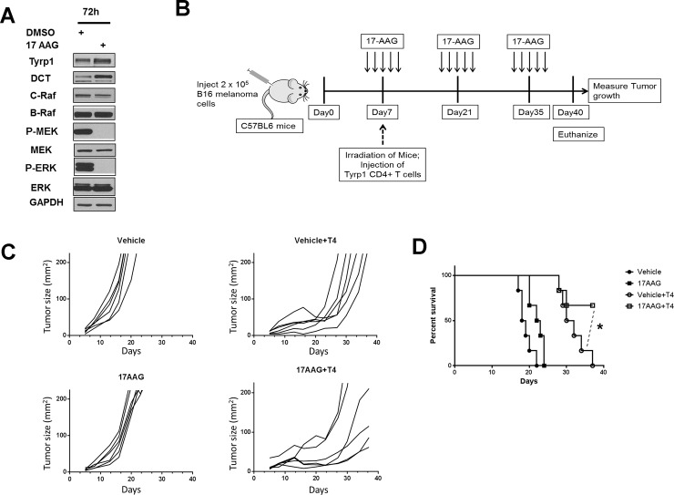
Fig 8. 17AAG promotes the inhibition of Tyrp1 specific CD4+ T cell treated melanoma tumor growth.
(A) Cultured B16 mouse melanoma cell lines were incubated with 17-AAG (1.0 μM) for 72 h, and cell lysates collected and studied for protein and phosphoprotein expression following SDS-PAGE and Western transfer. (B) The C57BL6 mice were subcutaneously injected with 2 x 10⁵ B16 mouse melanoma cells. Five days after B16 tumor cell injection, the mice received intraperitoneal injection of either 17AAG or vehicle at a dose of 75 mg/Kg body weight x 5 consecutive days. On seventh day after tumor challenge, all mice were irradiated one time with 550 rads (5.5Gy). 50% of 17AAG and 50% of Vehicle treated tumor bearing mice received 2 x 10⁵ numbers of TYRP1-specific CD4+ T cells (T4 cells) were injected intravenously. The 17-AAG dose of 75 mg/kg x 5 consecutive days was repeated every 2 weeks following initial administration of 17-AAG until the end of experiment. (C) The time dependent effect on the B16 melanoma tumor growth in C57BL6 mice treated either with Vehicle, 17AAG, Vehicle+T4 cells or 17AAG+T4 cells were determined by measuring their growth at regular intervals till the end of the experiment (n = 6). Data is a representative of two independent experiments. (D) The survival curve analysis shows that mice receiving no T4 cell transfer and only T4 cell transfer had no surviving mice at day 40. (*P<0.05 is a comparison between Vehicle+T4 cells and 17AAG+T4 cells as indicated by dotted lines).

