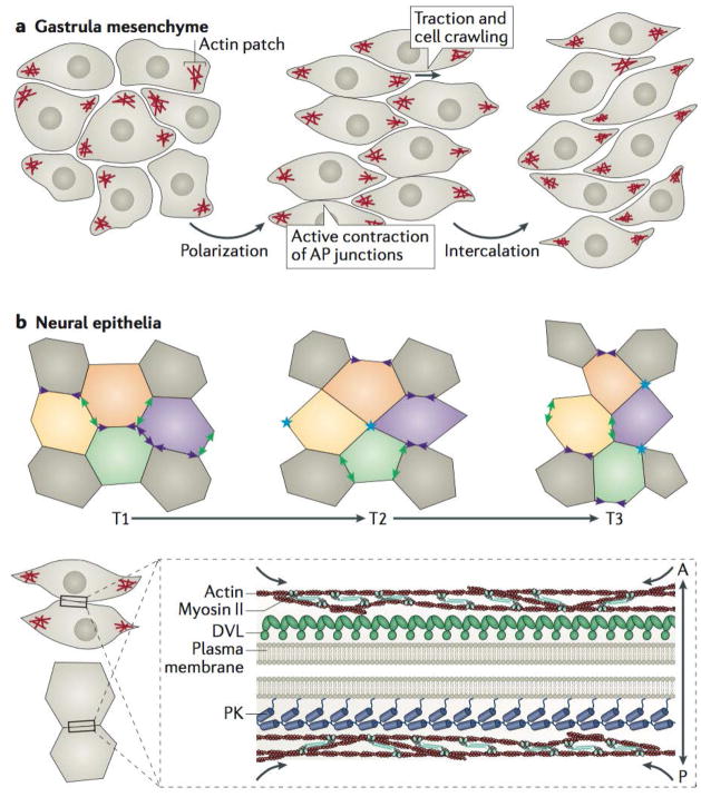
Figure 5. Planar cell polarity signalling directs polarized cell rearrangements during convergent extension.
a | Mesenchymal cells during vertebrate gastrulation collectively elongate along the mediolateral axis, stabilize mediolateral actin-based protrusions, and mediolaterally intercalate to narrow and lengthen the tissue; all these processes require intact planar cell polarity (PCP) signalling75,110. b | Neural ectodermal cells mediolaterally intercalate by preferentially shrinking antero-posterior (AP) junctions (purple arrowheads), which first resolve (blue asterisks) and then elongate to form new medio-lateral (ML) junctions (green arrowheads). The stages of this intercalation behaviour are often referred to as “T” transitions, where T1 to T2 transitions involve the shrinking of the AP junction, whereas T2 to T3 transitions involve the lengthening of a new ML junction between two cells previously separated along the ML axis at T1. c | Core PCP components can be found asymmetrically enriched along AP junctions of cells undergoing convergent extension in vertebrates, with Prickle (PK) typically localizing to the anterior cell faces and Dishevelled (DVL) along the posterior cell faces79–82. In the context of convergent extension, localization of PCP components at AP junctions coincides with the localization of filamentous actin enrichments and phosphorylated non-muscle Myosin II, which comprises the contractile actomyosin machinery that shrinks AP junctions81,99,101,102; there is evidence that PCP components are involved in regulating this contractility108,109.

