
Figure 2. The Vi Antigen Averts the Phagocyte Respiratory Burst by Inhibiting Antibody-Dependent Complement Activation.
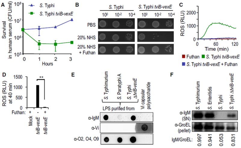
(A) Viability of the indicated bacterial strains incubated in 10% NHS was monitored over time.
(B) Viability of the indicated bacterial strains incubated in PBS, 20% NHS, or 20% NHS supplemented with Futhan was determined by spotting serial dilutions on agar plates.
(C and D) HL-60 cells were infected with the indicated opsonized bacterial strains and ROS production monitored over time using chemiluminescence.
(C) Representative experiment showing generation of chemiluminescence over time.
(D) Quantification of chemiluminescence from three independent experiments at the indicated time point after infection.
(E) Binding of IgM in human serum (α-IgM; top); binding of rabbit anti-Vi antigen serum (α-Vi; middle); or binding of rabbit anti-O2, -O4, -O9 serum (α-O2, O4, O9; bottom panel) to purified LPS of the indicated bacterial strains or to purified Vi antigen was detected by dot blot.
(F) The indicated bacterial strains were incubated in human serum and surface-bound proteins eluted with glycine. IgM eluted from the bacterial surface was detected by western blot (α-IgM; top). The bacterial protein GroEL was detected in the bacterial pellet (α-GroEL; bottom) and used as a loading control by measuring the IgM/GroEL using densitometry.
(A and D) Bars represent means ± SE.
Mock, mock infection; RLU, relative luminescence units. **p < 0.01.