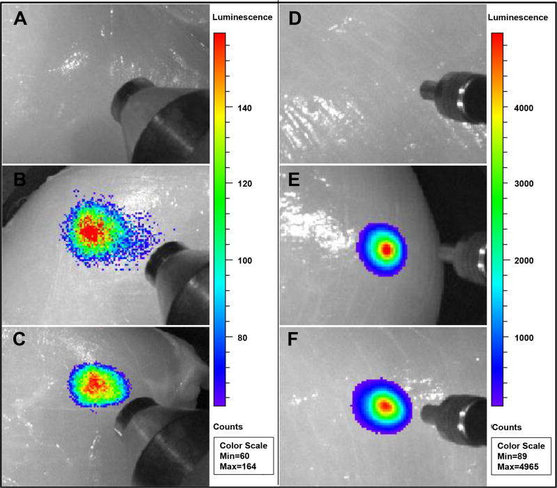
Figure 13.
Deep tissue imaging of X-ray or up-conversion nanophosphors: 200 µL of (A) PBS solution, (B) 1 mg/mL PEG-FA functionalized Gd2O2S:Eu (0% NaF doping), (C) 0.1 mg/mL PEG-FA functionalized Gd2O2S:Eu (7.6 mol% NaF doping), (D) PBS solution, (E) PEG-FA functionalized Y2O2S:Eu (0% NaF doping), and (F) PEG-FA functionalized Y2O2S:Eu (7.6 mol% NaF doping) were injected into chicken breast at a depth of 1 cm. A pseudocolor X-ray luminescence or upconversion luminescence intensity image was superimposed over a greyscale reflection image taken under an overhead lamp.

