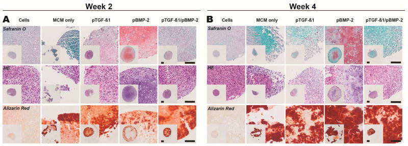
Figure 6.
Histologic characterization of Donor 1 MSC aggregates with incorporated microparticle-loaded transgenes. Tissue sections were stained with Safranin O for GAG (pink/red), hematoxylin and eosin, and Alizarin Red (red) for calcium after A) 2 and B) 4 weeks in vitro culture. HE, Hematoxylin and Eosin; MCM, mineral coated hydroxyapatite microparticles; TGFβ1, transforming growth factor-beta 1; BMP2, bone morphogenetic protein-2. Scale bars, 100 μm.

