Fig. 1.

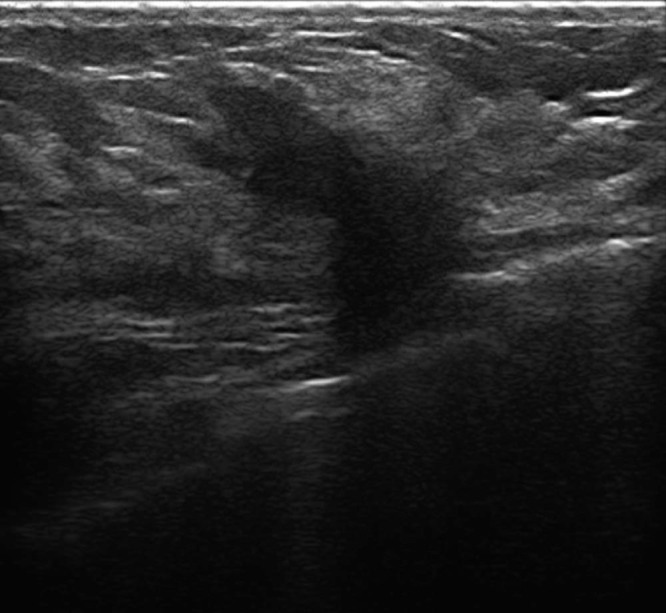
Ultrasound shows a 20-mm irregular, hypoechoic, solid mass with spiculated margins vascularized on color Doppler, with posterior acoustic shadowing in the inferior outer quadrant of the left breast.

Ultrasound shows a 20-mm irregular, hypoechoic, solid mass with spiculated margins vascularized on color Doppler, with posterior acoustic shadowing in the inferior outer quadrant of the left breast.