Fig. 5.

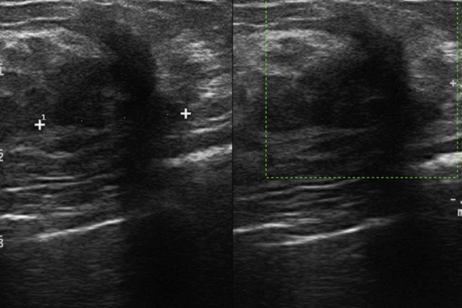
Inhomogeneously hypoechoic pseudonodular area of 23 mm, with ultrasound features and morphology similar to the lesion that had been surgically removed.

Inhomogeneously hypoechoic pseudonodular area of 23 mm, with ultrasound features and morphology similar to the lesion that had been surgically removed.