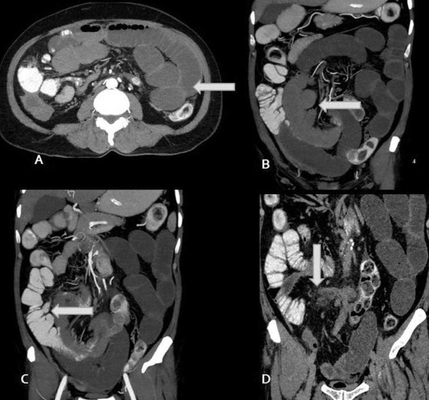
Fig. 2.
(A-D) Axial image (A) showing dilated jejunal loops (arrow) with coronal images (B-C) showing multiple variable-sized outpouchings from mesenteric side of dilated jejunal loops representing the diverticula (arrows) and last coronal image (D) showing inflammatory changes in the mesentery (arrow) in the form of high attenuation and marked stranding of mesenteric fat with small mesenteric lymph nodes. Adhesion band and adjoining thick narrowed small bowel jejunal loops can also be seen in the last image.

