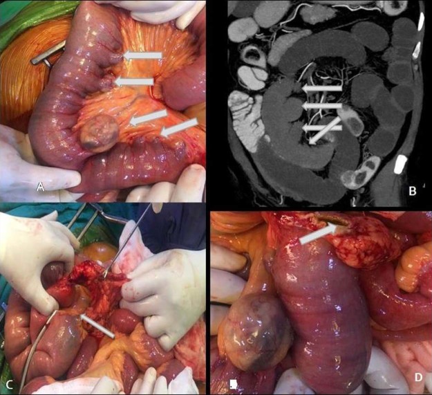
Fig. 5.
(A-D) Intraoperative image (A) shows multiple diverticula (arrows) arising from the mesenteric side of jejunal loops and almost corresponding coronal CT images (B) for comparison. Intraoperative image (C) shows adhesion band-forming tunnel (arrow) between mesentery and adhesion band with herniation of jejunal loops through this, and image D shows perforated diverticulum (arrow).

