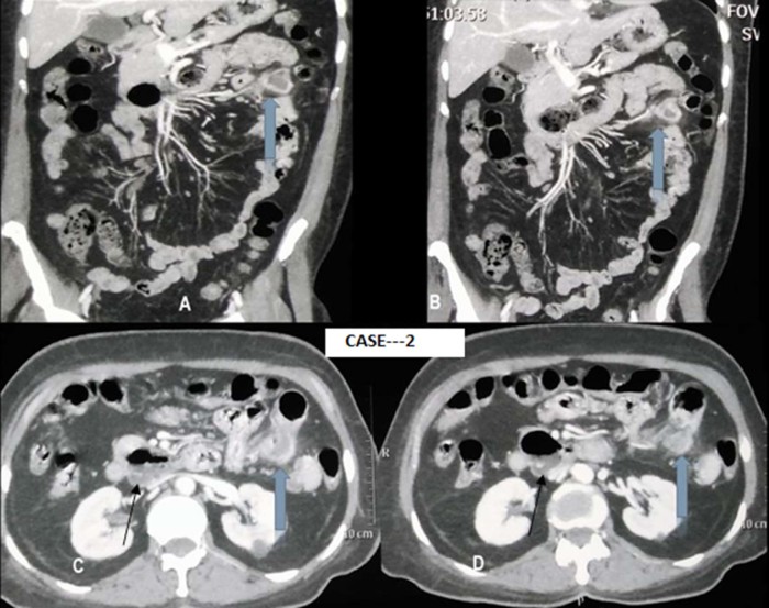
Fig. 6.
(A-D) CECT images show (wide arrows) inflamed jejunal diverticulum with formation of inflammatory mass showing peripheral enhancing wall with adjoining mesentery showing increased attenuation and significant stranding. Axial CECT image B (thin arrow) shows D3 duodenum diverticulum.
CECT, contrast-enhanced computed tomography.

