FIGURE 2.

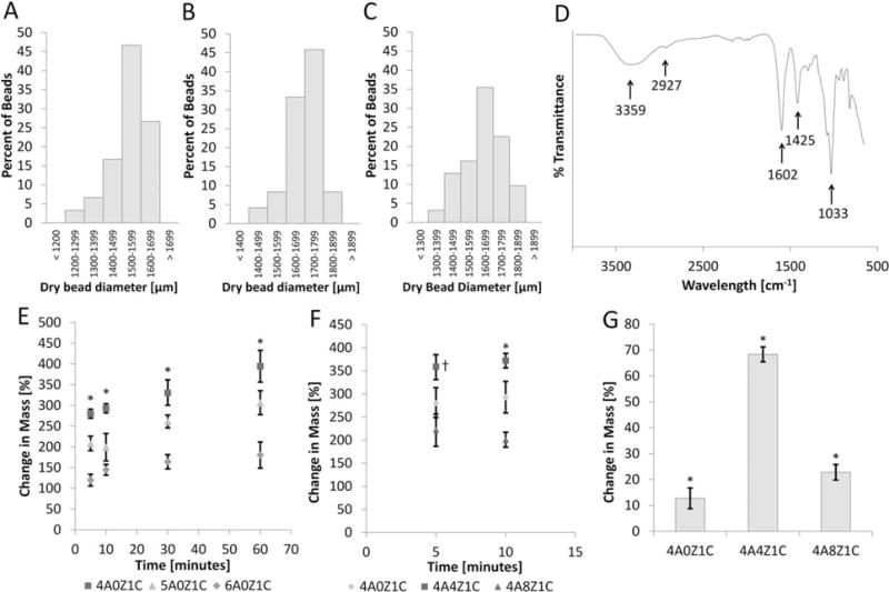
Sizing, FTIR, swelling, and mass loss. Bead sizes for (A) 4A0Z1C, (B) 4A4Z1C, and (C) 4A8Z1C are organized into bins with 100 μm ranges based on image analysis. (D) FTIR data for 4A4Z1C beads show absorbance peaks of N-H and O-H (broad band at ~3359 cm−1), O-H stretching (~2927 cm−1), C = O (~1602 cm−1), COOH (~1425 cm−1), SiO4 tetrahedral stretching (~1033 cm−1). (E) The average changes in mass of 4A0Z1C, 5A0Z1C, and 6A0Z1C beads are compared over one hour. Error bars show standard error. (F) The average changes in mass of 4A4Z1C and 4A8Z1C beads are compared at times pertinent to hemostasis. Error bars show standard error. (G) Mass loss in physiological conditions is assessed after 7 days. The error bar shows standard deviation. * indicates p < 0.05 compared within time points or between groups. † indicates p < 0.05 for group 4A4Z1C compared with both other groups.
