Figure.
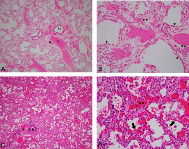
A-D, Histology from infants with classic and atypical presentations of ACDMPV. Hematoxylin and eosin–stained sections of lung with low-power (10×) and high-power (40×) views of classic ACDMPV (A, B) and atypical ACDMPV (C, D). Congested areas were selected for better visualization of the capillaries. A, Malposition of veins adjacent to small arteries within BVBs and lobular maldevelopment with decreased alveolarization. B, Almost complete absence of capillary loops adjacent to alveolar epithelium (*), with fewer and displaced capillaries in septae (**). C, Malposition of veins adjacent to small arteries in BVBs. Compared with the histology of classic ACDMPV, decreased alveolarization is not readily apparent. D, Areas of both normal capillarization with capillaries adjacent to alveolar epithelium (arrow), and deficient capillarization with displaced capillaries in septae (**). a, artery; b, bronchus; v, vein.

