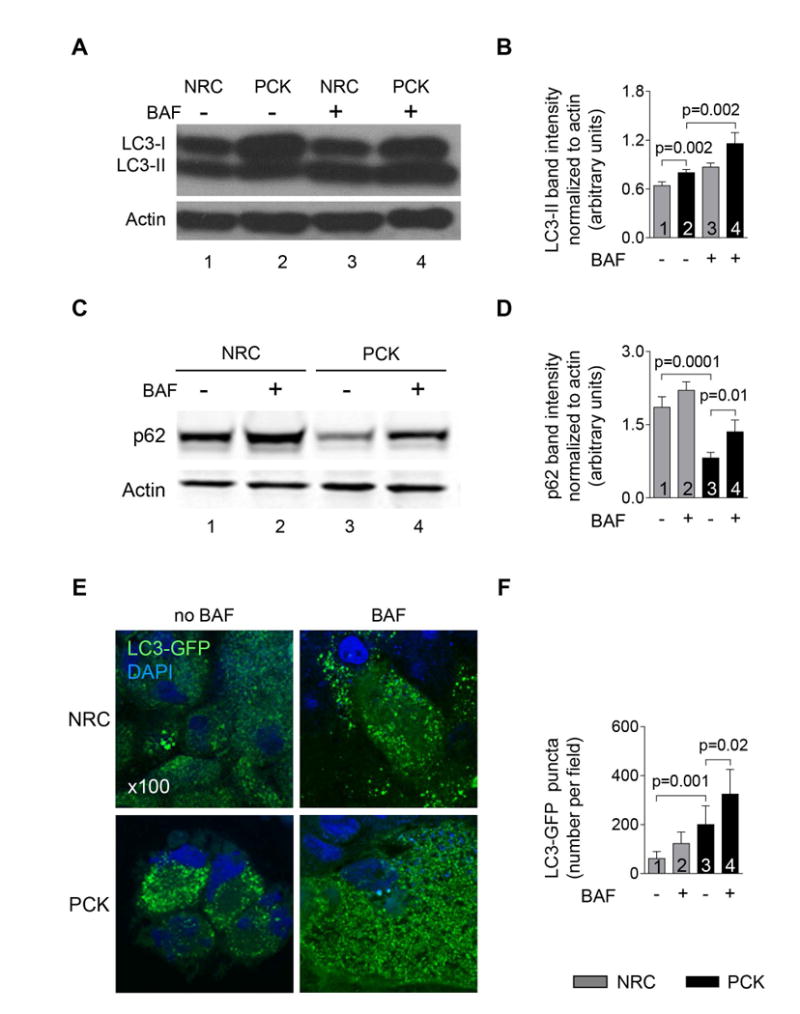
Figure 4. Autophagic flux is enhanced in PLD cholangiocytes.

(A&B) Expression of autophagosome marker, LC3-II, was greater in PCK rat cholangiocytes compared to NRC as demonstrated by representative western blots (line 2 versus line 1) and quantitative analysis of LC3-II band intensity (bar 2 versus bar 1). Bafilomycin A1 (100 nM) further increased LC3-II levels in PCK rat cholangiocytes (line 4 versus line 2; bar 4 versus bar 2) demonstrating enhanced autophagic flux. (C&D) Expression of p62/SQSTM1, an autophagy receptor, was lower in PCK rat cholangiocytes compared to NRC (line 3 versus line 1; bar 3 versus bar 1). Bafilomycin A1 (100 nM) increased p62 levels in PCK cholangiocytes (line 4 versus line 3; bar 4 versus bar 3) showing enhanced autophagic flux. (E) Representative immunofluorescence confocal microscopy images of NRC and PCK rat cholangiocytes stable transfected with LC3-GFP construct and (F) quantitation of LC3-GFP-positive puncta demonstrate that the number of LC3-GFP puncta is greater in PCK cholangiocytes compared to NRC (bar 3 versus bar 1). Treatment of PCK rat cholangiocytes with Bafilomycin A1 (100 nM) further increased the number of LC3-GFP-positive puncta (bar 4 versus bar 3) indicating increased autophagic flux. (n=3 for each cell line). Data are presented as MEAN±SD. Abbreviation: BAF – Bafilomycin A1.
