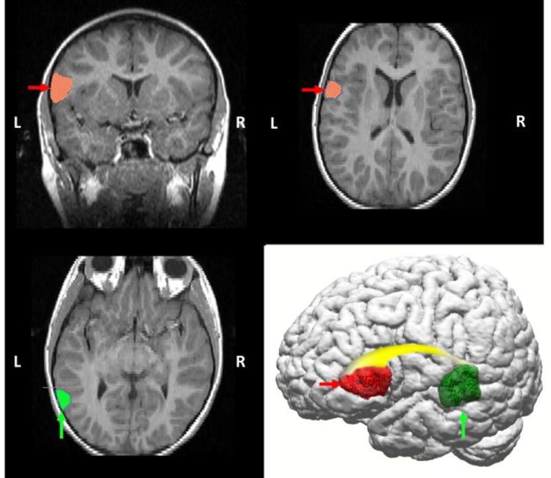
Figure 1.
Diagrammatic representation of primary language areas on anatomical T2-weighted FLAIR MRI and 3D reconstruction images (110), highlighting left temporal lobe (Wernicke’s area in posterior temporal region, vertical arrows/dotted hatching), frontal lobe (Broca’s area in middle frontal region, horizontal arrows/diagonal hatching), and the white matter (arcuate fasciculus, gradient shading) that connects these regions. Historically, language has been conceptualized as a lateralized function, with dominance typically in the left hemisphere. Functional magnetic resonance imaging (fMRI) studies confirm that language is a left-hemispheric dominant process in the vast majority of healthy adults (111), but also that language requires input from distributed networks, including homologous right hemispheric regions.

