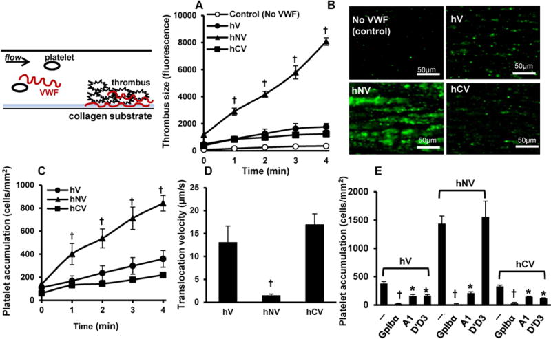
Figure 3. Thrombus formation and human platelet translocation.

A-B. 108/mL BCECF-labeled fluorescent human platelets were suspended in plasma-free blood supplemented with 10 μg/ml recombinant human VWF variants. The mixture was perfused over Type I collagen substrates for 4-5 min at a wall shear rate of 1000 s−1 (Shear stress ~40 dyn/cm2). A. Thrombus formation quantified based on total fluorescence intensity in the field of view. Control runs were plasma-free without addition of VWF. B. Representative thrombus images at 4 min for various cases. C-E. 108 washed platelets/mL were perfused over substrates bearing recombinant VWF variants for 4 min at a wall shear stress of 3 dyn/cm2. Both platelet accumulation (C) and translocation velocity (D) were recorded. E. In some cases, blocking mAbs were added at 20μg/mL to inhibit platelet binding. Data are mean + SD for 4min time point. Statistical symbols used are same as in Fig. 1. hV displays greater tendency to form thrombus.
