
Figure 1.
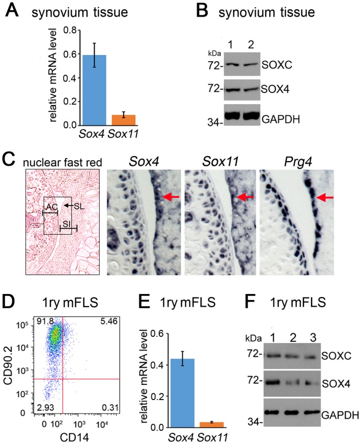
SOX4 and SOX11 are expressed in mouse fibroblast‐like synoviocytes (mFLS). A, Levels of Sox4 and Sox11 mRNA in synovial tissue from 6‐week‐old mice, as measured by quantitative reverse transcription–polymerase chain reaction and normalized to GAPDH mRNA levels. B, Expression of SOXC and SOX4 protein in whole synovium lysates from 2 wild‐type mice (mice 1 and 2). Western blots were hybridized with SOX4‐specific and pan–SOXC antibodies. GAPDH was used as the loading control. The molecular weights of protein standards running close to the proteins of interest are indicated. C, RNA in situ hybridization in adjacent knee sections from 6‐week‐old mice, using Sox4, Sox11, and Prg4 RNA probes. A nuclear fast red–stained section is shown in the left panel (original magnification × 5); the boxed area indicates the areas that are shown at higher magnification (original magnification × 20) in the middle and right panels. Arrows indicate synovial lining (SL). D, Flow cytometric phenotyping of mouse primary synovial cells at passage 4, using fluorescein isothiocyanate–conjugated CD90.2 and allophycocyanin‐conjugated CD14 antibodies. E, Levels of Sox4 and Sox11 mRNA in primary (1ry) mFLS similar to those in D. F, Detection of SOX4 and SOX11 proteins on Western blots of whole lysates from the immunophenotyped cells in D. Values in A and E are the mean ± SD. AC = articular cartilage; SI = synovial intima.