
Figure 4.
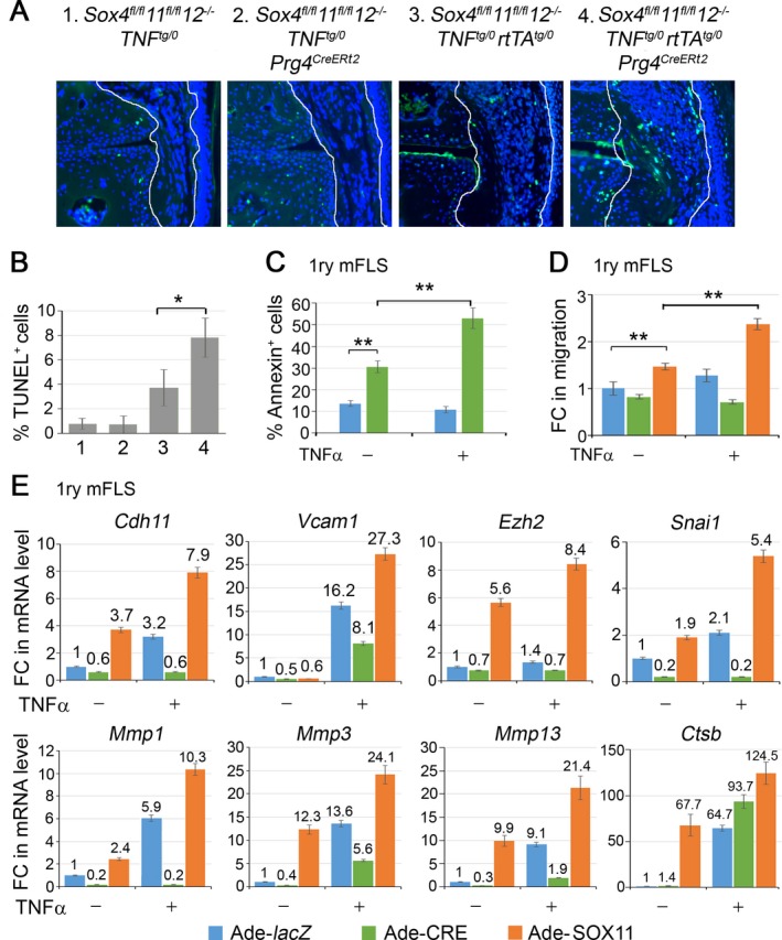
SOXC genes promote FLS transformation. A, TUNEL staining (green) and DAPI counterstaining (blue) of cell nuclei. Original magnification × 20. B, Death rate of cells in the synovium (n = 3 mice per genotype), as determined by TUNEL staining. C, Flow cytometric quantification of annexin V–positive cells in primary FLS from Sox4 fl/fl 11 fl/fl 12 –/– mice. Cells were infected with lacZ‐ or Cre‐expressing adenovirus for 16 hours and then treated with 5 ng/ml tumor necrosis factor (TNF) for 8 hours. D, Fold changes (FCs) in FLS migration to wounded area, as assessed by in vitro wound‐healing assay. Confluent cultures of FLS from Sox4 fl/fl 11 fl/fl 12 –/– mice were infected with lacZ‐, Cre‐, or SOX11‐expressing adenovirus (Ade) for 16 hours, a cell monolayer was scratched with a pipette tip, and the cultures were either treated with 5 ng/ml TNF for 4 hours or were left untreated. E, Fold changes in the levels of mRNA for genes required for efficient cell migration and FLS transformation. GAPDH mRNA levels were used for normalization. Values are the mean ± SD of triplicate cultures per condition. * = P < 0.05; ** = P < 0.01 by Student's 2‐tailed t‐test. See Figure 1 for other definitions.