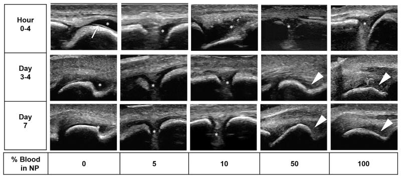
Figure 3. Time course of ultrasound appearance of blood dilutions and clotted blood in the MTP joints of cadaveric pig feet.
NP, diluted with increasing concentrations of freshly drawn human blood (3–5 mL) were injected into the MTP joint space. Serial ultrasound and sonopalpation was performed to identify fluid versus soft tissues at the indicated time points. Arrow: Interface between anechoic articular cartilage and fluid. Asterisk: Fluid-filled compressible areas in the joint space. Triangle : Partially or non- compressible clotted and aging blood products. Non-bloody fluid was anechoic. Echogenic signals could be appreciated starting as low as 5–10% blood dilution. Aging blood products/clots appeared hypoechoic and granular relative to the surrounding soft tissue. NP, Normal Pooled Plasma; MTP, Metatarsophalangeal.

