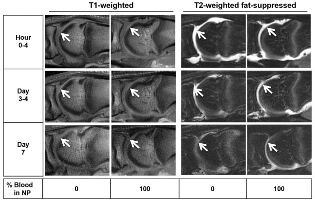
Figure 4. Time course of MRI appearance of blood dilutions and clotted blood in the MTP joints of cadaveric pig feet.
NP, diluted with increasing concentrations of freshly drawn human blood (3–5 mL) were injected into the MTP joint space. Serial MRI was performed using T1-weighted and T2-weighted fat- suppressed sequences at the indicated time points.. Arrows: Fluid-filled areas in the joint space. There was no appreciable difference in signal intensity between plasma and blood solutions on T1- and T2-weighted fat-suppressed sequences. NP, Normal Pooled Plasma; MRI, Magnetic Resonance Imaging; MTP, Metatarsophalangeal.

