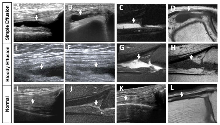
Figure 5. Timely correlated MSKUS and MR images in 2 hemophilic patients with simple and bloody effusions in relation to normal anatomy.
A/B: a 70-year-old-man with a simple effusion on MSKUS: Longitudinal image through the suprapatellar recess and short axis image through the lateral recess show anechoic joint fluid (arrows) consistent with simple effusion, confirmed through joint aspiration. E/F: a 57-year-old man with a bloody effusion on MSKUS: Longitudinal images through the suprapatellar recess applying different degrees of real-time compression show joint fluid with echogenic reflectors (arrows), consistent with bloody effusion, confirmed through joint aspiration. C/D and G/H: Corresponding MR images to A/B and E/F. Sagittal T2-weighted fat-suppressed and axial/sagittal T1-weighted images confirm joint effusions (arrows) in both cases, but are unable to distinguish between simple and bloody effusion. Simple fluid in C and D appears qualitatively similar to the bloody fluid in G and H. I/K and J/L represent normal sonoanatomy and MR anatomy of the suprapatellar and lateral recess, respectively. The normal recesses, which are entirely collapsed, are annotated with arrows. MSKUS, Musculoskeletal Ultrasound; MR, Magnetic Resonance.

