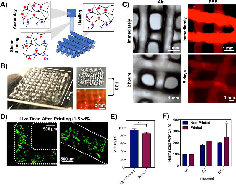
Figure 5. 3D printing of hydrogels into 3D, multilayer lattices.
A) Schematic of shear-thinning and self-healing of hydrogels during printing, where hydrogels were formed in syringes, sheared through the extrusion process, and filaments stabilized by self-healing properties of hydrazone bonds. B) Photos of 4-layer lattices in air and in PBS. C) Images of lattices in air and in PBS (labeled with rhodamine-dextran). D) Live/Dead staining of cells in lattice filaments immediately after extrusion and E) quantification of cell viability before and after printing based on Live/Dead staining. ***p<0.001. F) Metabolic activity of cells in hydrogels before and after printing by alamarBlue measurements of activity. *p<0.05 compared to D1.

