Fig. 1.
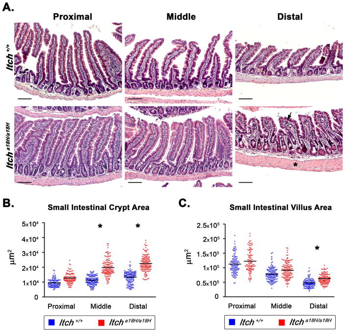
Small intestinal architecture is altered in Itcha18H/a18H animals. (A) Representative H & E stained paraffin-embedded small intestinal sections derived from the proximal (left most column), middle (center column), and distal (right hand column) segments of young adult Itch+/+ and Itcha18H/a18H animals accentuate an enlargement of the intestinal crypt, villus blunting with crowding of epithelial cells, and thickening of the muscularis propria (asterisk) in Itcha18H/a18H animals, becoming more pronounced distally. Mild inflammation was also occasionally noted in the lamina propria (arrow). Scale bars = 200 μm. (B) Small intestinal crypt and villus areas from young adult Itch+/+ (n = 4 or 5) and Itcha18H/a18H (n = 5) were measured in μm2 using ImageJ. A minimum of 25 well-orientated crypts or villi from at least seven different 10x fields were measured per animal. Average crypt area was significantly increased (*p < 0.005) in the middle and distal small intestine of Itcha18H/a18H compared to Itch+/+. (C) Average villus area was significantly increased (*p < 0.05) distally in the small intestine of Itcha18H/a18H animals compared to Itch+/+.

