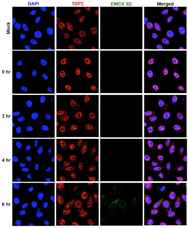
Fig. 1. TDP2 is re-localized from the nucleus to the cytoplasm during EMCV infection.
HeLa cells were seeded on coverslips and either mock- or EMCV-infected at an MOI of 20. Cells were then fixed with formaldehyde at 0, 2, 4, or 6 hr post-infection. Proteins were visualized by confocal microscopy using antibodies against human TDP2 (red) or EMCV 3D (green). Nuclei were counterstained with DAPI (blue).

