Figure 3.
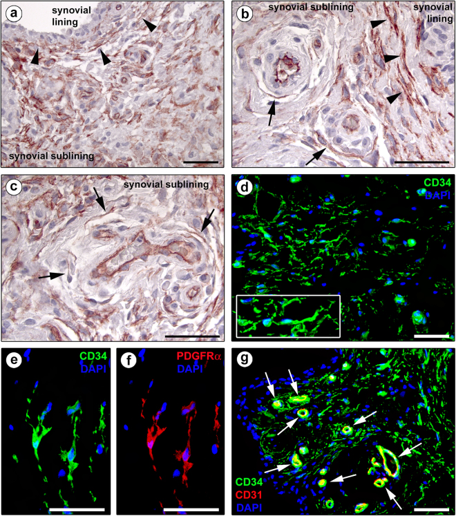
Representative light and fluorescence microscopy photomicrographs of human synovial sections. (a–c) CD34 immunoperoxidase-based immunohistochemistry with hematoxylin counterstain. (d) Immunofluorescence labeling for CD34 (green) with 4′,6-diamidino-2-phenylindole (DAPI; blue) counterstain for nuclei. CD34-positive spindle-shaped cells (telocytes) with long cytoplasmic processes (telopodes) are distributed throughout the whole synovial sublining layer (a–d). Note the presence of numerous CD34-positive stromal cells arranged in multiple parallel rows at the synovial lining-sublining interface (arrowheads in a,b) and surrounding sublining microvessels (arrows in b,c). CD34 immunopositivity is observed also in vascular endothelial cells. At higher magnification, the processes of CD34-positive stromal cells exhibit a moniliform silhouette (d, inset). (e,f) Double immunofluorescence labeling for CD34 (green) and PDGFRα (red) with DAPI (blue) counterstain for nuclei. Telocytes are double positive for CD34 and PDGFRα. (g) Double immunofluorescence labeling for CD34 (green) and CD31 (red) with DAPI (blue) counterstain for nuclei. Telocytes are CD34-positive/CD31-negative, while vascular endothelial cells are CD34-positive/CD31-positive (arrows). Scale bar: 50 µm (a–g).

