Figure 4.
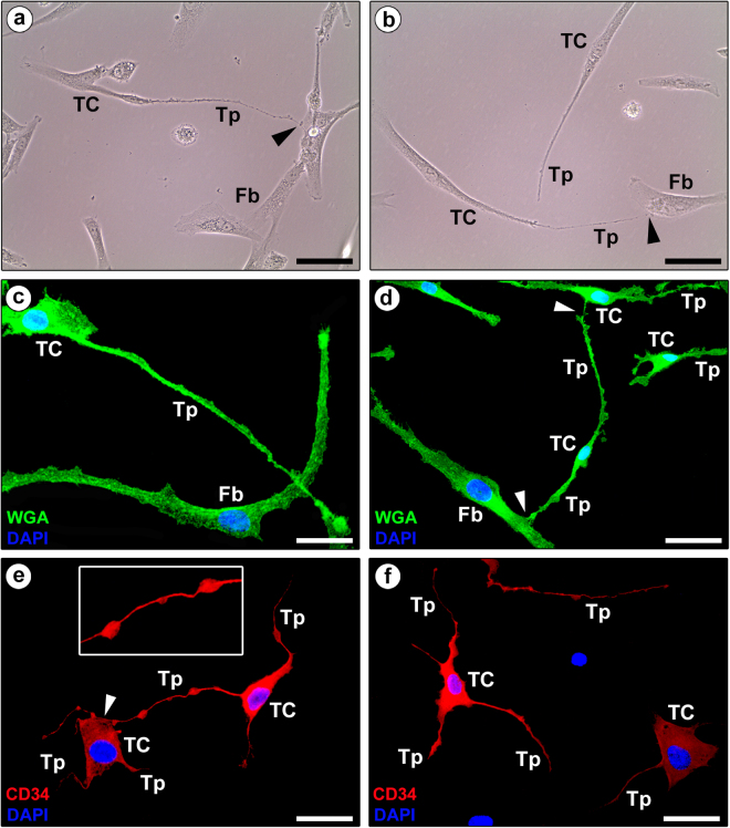
Representative phase-contrast and fluorescence microscopy photomicrographs of human fibroblast-like synoviocyte primary cultures. (a,b) Phase-contrast microscopy. (c,d) Wheat Germ Agglutinin (WGA; green) immunofluorescence labeling with 4′,6-diamidino-2-phenylindole (DAPI; blue) counterstain for nuclei. (a–d) Note the presence of either fibroblasts (Fb) or telocytes (TC), the latter being characterized by a small cell body and very long and thin moniliform processes (telopodes, Tp) often establishing intercellular contacts with fibroblasts or other telocytes (arrowheads). (e,f) Immunofluorescence labeling for CD34 (red) with DAPI (blue) counterstain for nuclei. CD34-positive cells projecting very long and thin moniliform cytoplasmic processes (telopodes, Tp) are unequivocally identifiable as telocytes (TC). The inset in (e) shows a higher magnification of a typical telopode formed by an alternation of thin segments (podomers) and expanded parts (podoms). Note the telopode of a telocyte contacting the cell body of another telocyte (arrowhead in e). Scale bar: 50 µm (a–f).

