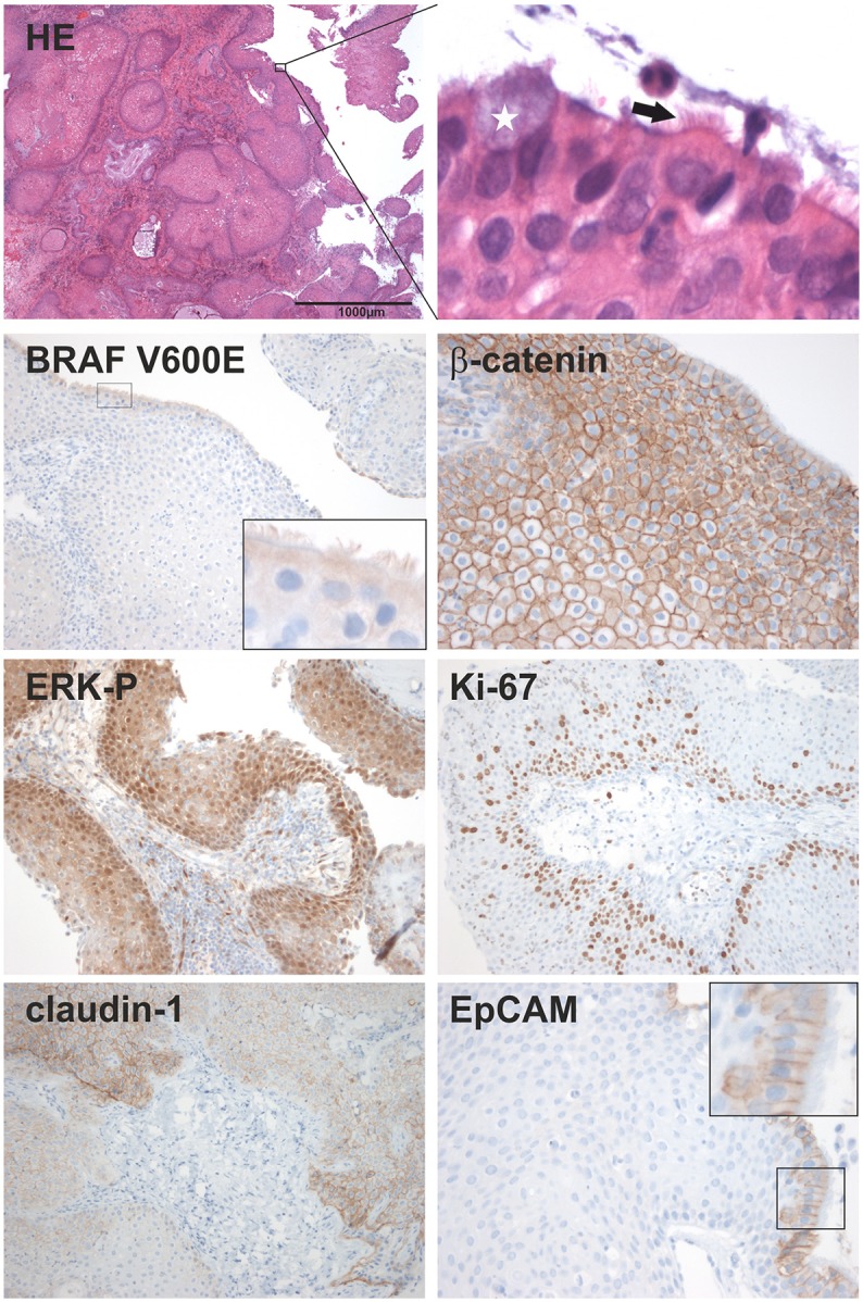
Figure 2.

Histological characterization; HE staining shows a lesion of squamous couple stone differentiated epithelium. Magnification revealed a superficial cell layer harboring kinocilia (arrow) and goblet cells (white star). The following immunohistochemical stainings show the evidence and expression pattern of the mutated BRAF V600E protein, beta-catenin; the phosphorylated ERK (ERK-P) and the proliferation marker Ki-67; as well as the distribution of the cell-cell contact proteins, claudin-1 and EpCAM. In a higher resolution, EpCAM is only detectable in ciliated cells of the superficial cell layer.
