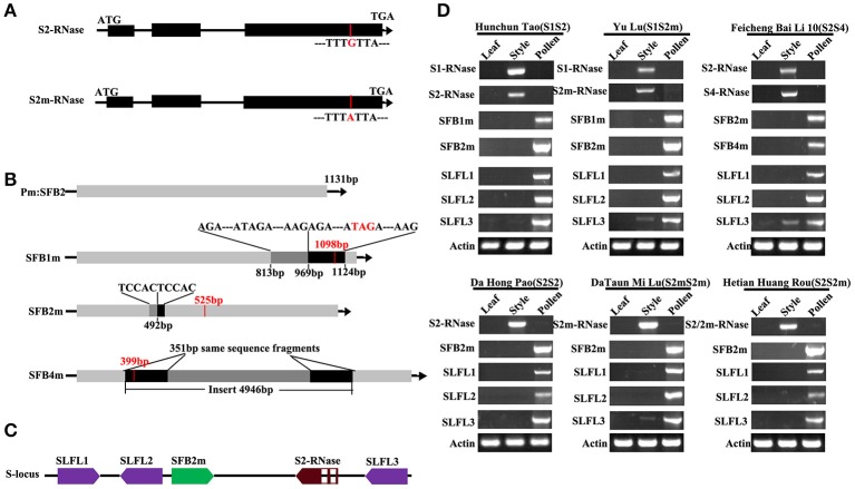
Figure 1.
Characterization and expression patterns of peach S genes. (A) Schematic diagrams of the S2/2m-RNase. The red line represents the mutation site of the S2-RNase. (B) Schematic diagram of peach PperSFBs. The black arrows indicate the transcriptional orientations of the genes. The red vertical bars indicate the stop codon, and the red numbers represent the length of the encoding frame. The black boxes in PperSFB1m and PperSFB2m represent the inserted fragment, and the gray boxes represent the same fragment of the gene as the inserted fragment. The black boxes in PperSFB4m represent the same fragments at both ends of the inserted fragment. (C) Schematic diagrams of the location of PperS2-RNase, PperSFB2m, and PperSLFLs at the S-locus. The directions of the arrows represent the transcriptional orientations of PperS2-RNase, PperSFB2m, and PperSLFLs. The middle parts in the red box represent the introns. (D) Tissue-specific expression analysis of PperS1-RNase, PperS2-RNase, PperS2m-RNase and PperS4-RNase, PperSFB1m, PperSFB2m, PperSFB4m, and PperSLFLs. Total RNA from different organs was extracted and used as template for cDNA synthesis.

