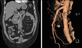
Figure 1.

Coronal reformatted image from contrast enhanced computed tomography demonstrating small pseudoaneurysm related to the superior aspect of a mesenteric root haematoma (left); 3D reconstruction showing pseudoaneurysm of inferior pancreaticoduodenal artery (right)
