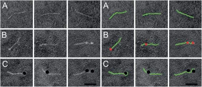
Figure 2.
Negative staining electron microscopy of SIC in complex with histone H4. Right panel shows a duplicate of the left panel, but with SIC (green) and histone H4 (red) illustrated in pseudocolors. (A) Linear SIC monomers in the absence of a ligand. (B) Histone H4 was added to SIC and prepared for negative staining electron microscopy. SIC monomers are depicted with one or two bound molecules of histone H4. (C) Histone H4 was labeled with 10-nm colloidal gold and added to SIC. SIC monomers binds to one or two molecules of histone H4 conjugated to gold particles. Scale bar 25 nm.

