Figure 1.
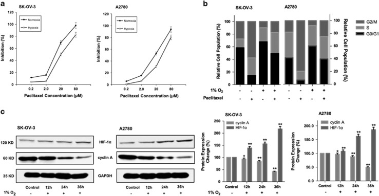
Hypoxia deteriorated G2/M arrest induced by paclitaxel. (a) Cell growth inhibition of paclitaxel treatment under normoxia and hypoxia assessed by MTT assay. Data had been statistically analyzed by Microsoft Excel 2013 and expressed as means±S.D. for three independent experiments. (b) The influence of paclitaxel treatment on cell cycle under normoxia and hypoxia. Summary of the percentage of cells at G0/G1, S and G2/M phase was performed. (c) After 12, 24 and 36 h hypoxic incubation, cyclin A and HIF-1α expression detected by western blottings. Protein expression change was represented by densitometric analysis. The results are representative of three independent experiments and expressed as means±S.D., *P<0.05 and **P<0.01, compared with the control groups

