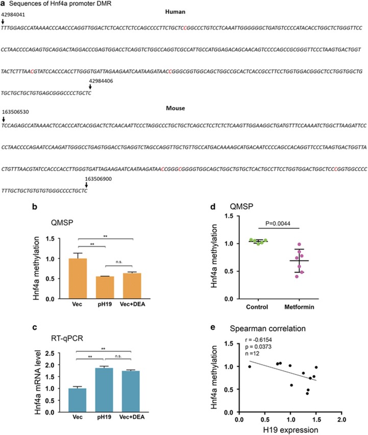
Figure 4.
H19 regulates Hnf4α methylation in vitro and in vivo. (a) Sequences of DMRs in the conserved promoter region of human and mouse Hnf4α. The three differentially methylated cytosine residues are highlighted in red. The numbers on top of the sequences mark the positions of the indicated nucleotides in the chromosomes. (b) Huh7 cells were transfected with Vec, pH19, or Vec plus DEA. Genomic DNAs were extracted 15 h later and analyzed by QMSP. Numbers are mean±S.D. (n=3). **P<0.01. NS, not statistically significant. (c) Huh7 cells were treated as described in b. RNAs were extracted 24 h later and analyzed by RT-qPCR. Numbers are mean±S.D. (n=3). **P<0.01. NS, not statistically significant. (d) Scatter plot of Hnf4α methylation in mouse fetal livers. Control group, n (fetuses from 5 dams)=5; metformin group, n(fetuses from 7 dams)=7. (e) Spearman correlation between H19 RNA level and Hnf4α promoter methylation, showing a negative correlation

