Fig. 3.
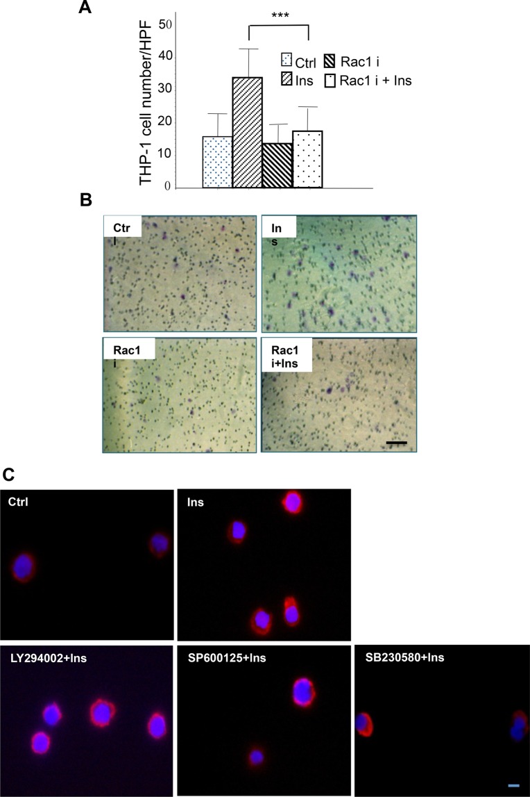
Insulin-induced Rac1 activation is regulated by PI3K-Akt and SPAK/JNK signal but not p38. (A,B) THP-1 cells were pre-treated with 50 μM Rac1 inhibitor NSC23766 for 30 min, chemotaxis assay was then performed with or without 10−7 M insulin as described in Fig. 1. Insulin-induced chemotaxis was inhibited by Rac1 inhibitor. ***P<0.001, n=3. Statistical analysis was performed as described in the Materials and Methods section; data are shown as mean±s.d. (B) Represented pictures of chemotaxis of THP-1 Cells. (C) Cells were pre-treated with 50 μM PI3K inhibitor LY294002, 50 μM SPAK/JNK inhibitor SP600125 and 10 μM P38 inhibitor SB203580 for 1 h, followed by treatment with 10−7 M insulin for 15 min. Cells were then collected and fixed, and were stained using Rac1–TRITC antibody. Rac1 distribution was visualized by immunofluorescence microscopy. Insulin-induced Rac1 distributed to the leading edge of migrating cells, which could be inhibited by PI3k and SPAK/JNK inhibitors but not P38 inhibitors. C is representative of three different experiments. Scale bar: 10 µm.

