
Fig. 1.
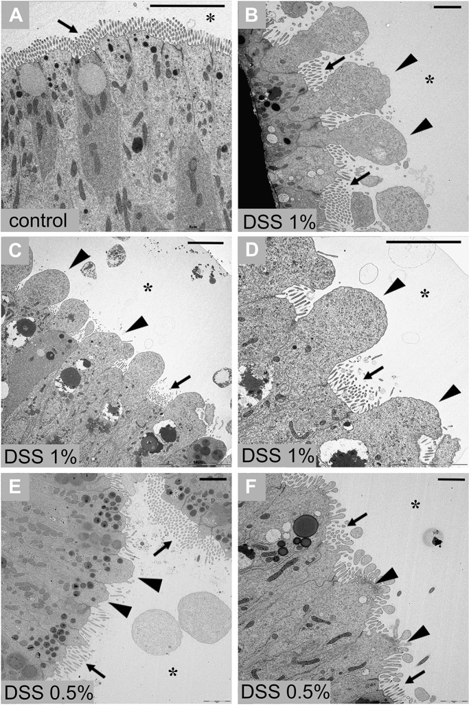
TEM of stomach epithelium of adult Ciona. Stomach epithelium of control animal, not treated with DSS (A). Stomach epithelium of animals treated with 1% DSS (B-D) and 0.5% DSS (E-F) exhibits morphological alterations that include reduction or loss of the microvillar structures and cytoplasmic extroflession in the areas facing the lumen (asterisk). n=8 animals, collected from the wild, observed for each condition. Arrows indicate microvilli of epithelium; triangles indicate cytoplasmic extroflession. Scale bars: 5 µm.