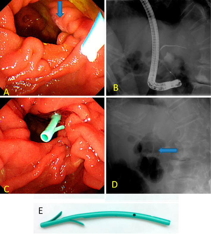
Figure 2.
Endoscopic (A, C) and fluoroscopic (B, D) images during endoscopic retrograde cholangiopancreatography. (A) The papillary orifice (blue arrow) was seen at the right side of the diverticular rim. (B) Selective bile duct cannulation was unsuccessful, although pancreatic guide-wire cannulation was performed simultaneously. (C, D) A pancreatic spontaneous dislodgement stent (blue arrow) was inserted to prevent post-ERCP pancreatitis. (E) The stent used was a polyethylene 5F diameter, 3 cm in length, straight-type stent, unflanged on the pancreatic ductal side with 2 flanges on the duodenal side (GPDS-5-3; Cook Japan, Tokyo, Japan).

