Figure 1.

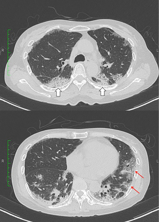
CT scan of the chest on admission showing nonsegmental consolidation (void arrows) and ground-glass attenuation (arrows) in the bilateral lung fields.

CT scan of the chest on admission showing nonsegmental consolidation (void arrows) and ground-glass attenuation (arrows) in the bilateral lung fields.