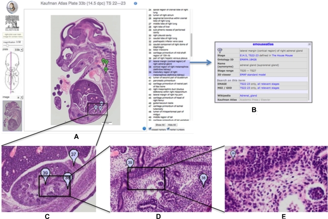
Figure 1:
Kaufman Atlas eHistology Viewer. A, Screen shot showing the user interface with annotation list. The 3 selected terms showing as a blue “flags” numbers 37, 38, 39 are over the developing kidney. B, The pop-up dialog with extra information on selecting the “37.” C–E, Progressively higher-resolution images corresponding to zooming-in on the image. At full resolution, the pixel spacing is 0.34 × 0.34 microns and reveals cellular architecture and arrangements.

