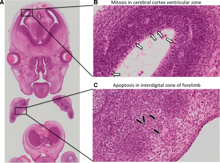
Figure 2:
Observing mitosis and apoptosis in cellular resolution eHistology atlas images. A key advantage of capturing histology images at high resolution is the ability to morphologically identify mitotically dividing cells and apoptotic cells in embryo atlas images. A, Zoomed-out view of a coronal image of an E14.5 embryo. B, On the zoomed-in view, neuroblasts in the ventricular zone of the cerebral cortex show intense haematoxylin staining (white arrows), a morphological feature associated with chromosome condensation in mitotically dividing cells. C, On the zoomed-in view, scattered cells in the interdigital zone of the forelimb are pyknotic (black arrows), a morphological feature associated with nuclear condensation. The pyknotic cells additionally show signs of cell shrinkage. These are morphological hallmarks of apoptosis.

