FIG 4.
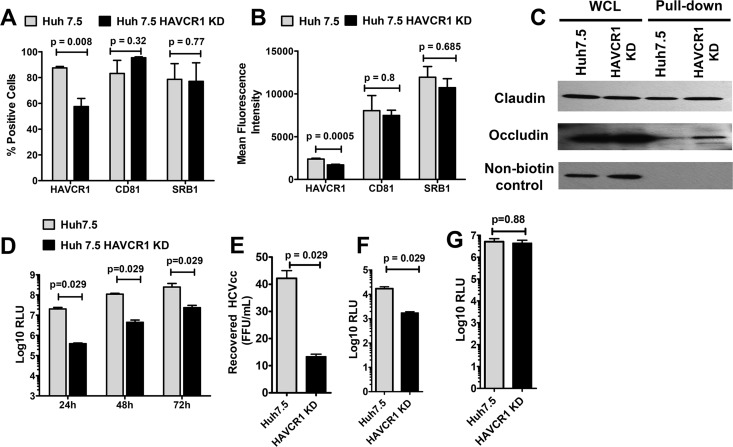
Reduced HAVCR1 expression on Huh7.5 cells results in reduced levels of HCVcc and HCVpp entry. Huh7.5 HAVCR1-1 knockdown (KD) cells were generated by transfection of parent Huh7.5 cells with a plasmid expressing HAVCR1 shRNA. (A) Percentages of parent and KD cells positive for HAVCR1, CD81, and SRB1 surface expression. Bars represent the mean values from 5 independent experiments for HAVCR1 and CD81 and 4 independent experiments for SRB1. (B) Mean fluorescence signals of HAVCR1, CD81, and SRB1 surface expression on parent and KD cells. Bars represent the mean values from 5 independent experiments for HAVCR1 and CD81 and 4 independent experiments for SRB1. (C) Western blot detection of claudin and occludin after cell surface biotinylation of Huh7.5 and HAVCR1 KD cells. Whole-cell lysate (WCL) is cell lysate prior to pulldown treatment. Nonbiotinylated control represents cells treated exactly as for the biotinylated cells but without the addition of EZ-Link sulfo-NHS-LCLC-biotin for cell surface biotinylation. Anti-claudin antibody was used for detection and shows the specificity of the NeutrAvidin beads for precipitation. (D) Luciferase activities at 24, 48, and 72 h postinfection in lysates of parent and KD cells infected with JFH1-Nanoluc. Bars represent the mean relative light units (RLU) calculated from 4 independent experiments using 9 replicates per experiment. (E) Titers of virus recovered from the supernatants of parent and KD cells at 72 h postinfection with JFH1-Nanoluc. Bars represent the mean titers from 4 independent experiments. (F) Luciferase activities in parent and KD cells 48 h after infection with HCVpp. (G) Luciferase activities in parent and KD cells 48 h after infection with VSV-G. Bars represent the mean values from 4 independent experiments. Error bars in all graphs represent standard errors of the means. Statistical analyses were performed using the Mann-Whitney test.

